
[ad_1]
Constructing an atlas of all of the cell varieties that make up the physique usually requires multinational collaborations and large budgets. However a method that may analyse the genetic exercise of a whole lot of 1000’s of particular person cells at a time has allowed one small crew to provide a time-lapse atlas of an embryonic mouse’s cells over ten days of improvement. The atlas, which was created by three researchers in a single 12 months for roughly US$370,000, may assist scientists to know how stem cells flip into particular cell varieties, how organs develop and even how the physique adjustments simply after it’s born.
The research, which was revealed in Nature in February1, “is spectacular at many ranges, each the size of what they achieved and the way they achieved it”, says Bertie Göttgens, a stem-cell biologist on the College of Cambridge, UK, who was not concerned within the research.
Geneticist Jay Shendure on the College of Washington in Seattle doesn’t usually research mouse improvement. His laboratory is understood for establishing molecular-biology strategies, together with one referred to as sci-RNA-seq3 that enables researchers to survey the assemblage of messenger RNA (mRNA), recognized collectively because the transcriptome, in particular person cells.

Which single-cell evaluation instrument is finest? Scientists provide recommendation
As a substitute of entire cells, which might be tough to maintain intact by way of the method, scientists grind up a pattern — on this case, an entire mouse embryo — and isolate its cell nuclei. They break up these nuclei into particular person dishes and add a distinct molecular tag to the mRNA in every dish. Subsequent, they mix the nuclei, separate them once more, mark every dish with a brand new tag and repeat. Ultimately, every nucleus acquires a singular assortment of tags — a molecular barcode — that the researchers can use to find out which tags outline the cell’s transcriptome. They will then sequence these cells’ mRNA and assemble a ‘tree’ that fashions how one cell kind can flip into one other, doing so throughout a number of animals of various ages on the idea of the genes they categorical.
Lacking moments
Two of Shendure’s lab members, postdoc Chengxiang Qiu and analysis scientist Beth Martin, determined to reveal sci-RNA-seq3 by charting the single-cell transcriptomes of embryonic mice throughout the animals’ roughly 19-day gestation interval. At first, they collected embryos each 24 hours over a 5-day interval, however the transcriptomes modified a lot between time factors that it was tough to comply with how stem cells was particular cell varieties over time2. Shendure likens it to a video that’s lacking too many frames: extra like a stop-motion animation than a easy development.
So Martin and Qiu partnered with analysis scientist Ian Welsh on the Jackson Laboratory, a analysis institute and mouse-breeding facility in Bar Harbor, Maine. Welsh painstakingly collected 83 mouse embryos at 2–6-hour intervals over 10 days of gestation, from the purpose at which organs begin to develop up till simply after the animal’s beginning. Welsh snap-froze the embryos and despatched them to Seattle, the place Martin collected single-cell transcriptomes. Qiu then mapped the info into timber that show when and the way every of 190 cell varieties — liver or bone-marrow cells, as an illustration — originates in an embryo.

Good software program untangles gene regulation in cells
To flesh out the tree, the researchers built-in their knowledge, which started eight days into gestation, with current work from Shendure’s crew and others that had mapped the transcriptomes of those and youthful embryos. This added one other 110,000 cells to the combination, and these knowledge shaped the tree’s ‘roots’, permitting the researchers to comply with the branching of early stem cells into particular varieties seen within the older embryos.
The ensuing atlas, containing the transcriptomes of mice throughout 45 time factors, is now obtainable for developmental biologists to check in additional depth. With 12.4 million cells, it’s the largest mouse-embryo atlas up to now and is almost one-quarter the scale of the cell knowledge collected by the Human Cell Atlas collaboration, which contains 700 labs trying to map all the cells within the human physique.
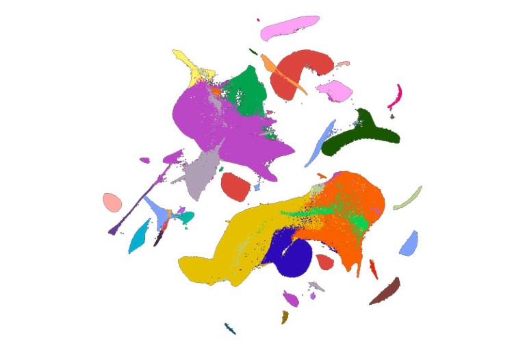
A 2D visualization of the mouse-atlas knowledge set, with colors equivalent to 26 main cell varieties.Credit score: C. Qiu et al./Nature
“It’s a improbable useful resource for the group,” says mobile geneticist and Human Cell Atlas co-founder Sarah Teichmann on the Wellcome Sanger Institute in Hinxton, UK. Teichmann factors out that there’s nonetheless work to be accomplished on the mouse atlas. A while factors have extra full transcriptomes than others, and the researchers haven’t but separated mice by intercourse to take a look at these variations. However she says it can allow numerous research, together with the flexibility to check mouse and human improvement. Shendure says he and his crew plan to create single-cell atlases of juvenile and grownup mice from conception to loss of life.
Stress results
Though Shendure and his group goal to let others conduct in-depth organic analyses of the info, they did observe two phenomena of their paper. The purpose at which the transcriptome modified most dramatically, they discovered, was within the hour simply after beginning, which Shendure calls “essentially the most tense second in your life”. A few of these variations had been anticipated — lung and fats cells modified exercise to deal with being outdoors the uterus, as an illustration — however different adjustments are nonetheless unclear.

How single-cell multi-omics builds relationships
Pure luck led them to a different discovering. To get the timing excellent, Welsh usually delivered the mice by caesarean part. However in the future, he returned from lunch to an sudden nest of new child pups. Martin processed the mice anyway and located that their transcriptomes had been considerably completely different from these of mice born by caesarean part. These variations may clarify the variation in well being outcomes seen between individuals who had been born by these two strategies, the researchers say.
Yonatan Stelzer, an epigeneticist on the Weizmann Institute of Science in Rehovot, Israel, says the research is encouraging for future efforts to map the cells of particular person organs or tissues. The following step for embryos, he says, will contain not solely learning how cells develop over time, but additionally following them by way of house in 3D, monitoring how they break up and transfer to type an entire mouse. Future analysis, he provides, may additionally examine questions corresponding to how two cells with related transcriptomes find yourself with completely different fates to turn out to be the proper or left eye, as an illustration. “We’re nonetheless removed from fixing your entire embryonic puzzle,” he says.
[ad_2]
Supply hyperlink

